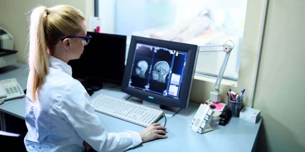
The Most Common Types of Medical Imaging

In the world of healthcare, imaging has revolutionized the field of medicine by providing non-invasive ways to visualize the body. It has allowed doctors to diagnose conditions faster and provide more accurate treatment. However, there is more than one type of medical imaging, each for different purposes depending on the specific medical condition and the part of the body being examined. So, let’s go over the different types of medical imaging.
The Different Types of Medical Imaging
1. Computed Radiography (CR) / Digital X-ray (DX)
Computed Radiography and Digital X-ray are two terms that are commonly used to refer to the same technology. CR is an older term that specifically refers to the use of a special plate to capture X-ray images, while DX refers to the use of a digital detector to capture X-ray images directly. In modern healthcare, CR/DX has largely replaced traditional film X-ray imaging—offering higher quality images, faster image capture, and more efficient image storage.
2. Computed Tomography (CT)
CT scans, also known as CAT scans, use ionizing radiation to produce multiple images of a body part, which are then combined to create detailed cross-sectional images of the body. This allows doctors to see organs and tissues from multiple angles. CT scans are commonly used to diagnose a range of conditions: cancers, brain injuries, cardiovascular disease, and other abnormalities. It can also be used to guide biopsies and plan surgeries.
3. Magnetic Resonance Imaging (MRI)
MRI uses a strong magnetic field and radio waves to produce detailed cross-sectional images of internal body structures. Unlike CT scans, MRI scans do not use ionizing radiation; instead, they use the magnetic properties of atoms in the body to create images. MRI can be used to diagnose medical conditions, including brain and spinal cord injuries, joint and muscle disorders, and some cancers.
4. Ultrasound (US)
Ultrasound, sometimes called sonography, uses high-frequency sound waves to create images. It is safe, non-invasive, and does not use ionizing radiation. This medical imaging technology is often used to image internal organs such as the heart, liver, and kidneys. It is also frequently used during pregnancy to monitor fetal development and check for any abnormalities.
5. Mammograms (MG)
A mammogram is used to examine the breast tissue for any abnormalities, including early signs of breast cancer. During a mammogram, the breast is compressed between two plates, and X-rays are taken from different angles to create a 2D or 3D image of the breast tissue. Mammography uses ionizing radiation; however, the amount of radiation exposure is typically very low.
6. Nuclear Medicine Imaging (NM)
Nuclear Medicine Imaging uses small amounts of radioactive materials—radiopharmaceuticals—as a tracer to produce images of the body. After the radiopharmaceutical is administered to the patient, a special camera is used to detect the radiation emitted by the tracer and create images of the body based on the distribution.
*Note: There are many other types of medical images. These are just a few.
The Integration of PACS Technology
PACS stands for Picture Archiving and Communication System. It is a technology used to store, retrieve, distribute, and display medical images. This platform can be used with a variety of imaging types, including the ones mentioned above and many more.
PACS allows medical professionals to view and analyze images from different types of medical imaging equipment, all in one place. This improves efficiency and accuracy in diagnosis and treatment planning, as medical professionals can access and easily compare images from different sources. By enabling the seamless integration of PACS into your healthcare environment, you can improve patient outcomes and enhance your overall healthcare delivery. For more than 25 years, Aspyra has been delivering complete PACS solutions to healthcare organizations. Contact us to learn more about what we can do for you.

